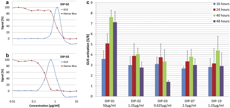
Figure 2. GUS differentiation signal is time- and concentration-dependent and coincides with cell death.
Serial dilutions of DIP-02 (a) and DIP-03 (b) in the GUS differentiation assay and the Alamar blue viability assay (40 h exposure time). GUS activation (S/B) of DIP-01, DIP-02, DIP-03, DIP-07 and DIP-19 at different exposure times (16 h, 24 h, 40 h and 48 h) at the concentration for optimal GUS expression (c). Bars indicate standard deviations of S/B from at least three independent assays. CA = cis-aconitate.

