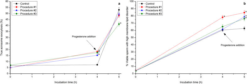
Figure 4. Percentages of true acrosome exocytosis and viable spermatozoa exhibiting high membrane lipid disorder following ‘in vitro’ capacitation and subsequent progesterone-induced ‘in vitro’ acrosome exocytosis after photo-stimulation procedures.
Boar sperm were subjected to separate photo-stimulation procedures and the subsequent IVC/IVAE as described in the Methods section. At the indicated times, aliquots were taken and percentages of true acrosome exocytosis (a) and of those viable sperm exhibiting high membrane lipid disorder (b; percentages of M-540+-sperm are shown considering viable sperm only) were determined. 0: values at 0 h of incubation in the capacitation medium (CM). 4: values at 4 h of incubation in the CM. 5: values after 4 h IVC plus 60 min of the progesterone-induced IVAE. Results are shown as mean ± SEM for 12 separate experiments. Asterisks indicate significant (P < 0.05) differences when compared with the respective Control value at the same time point.

