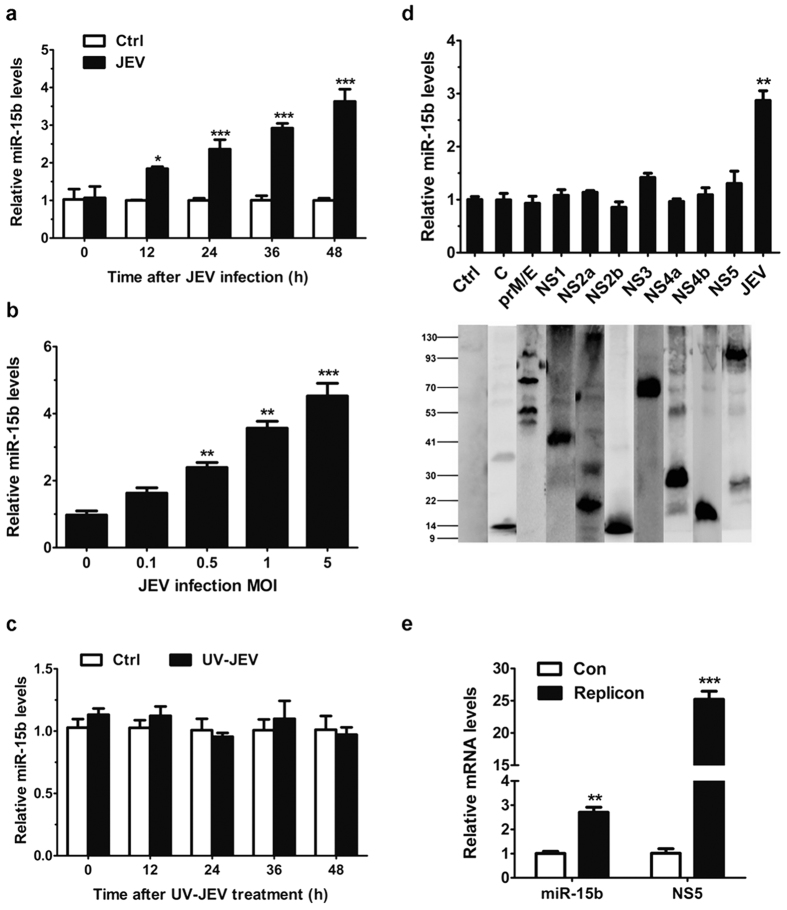
Figure 1. miR-15b is upregulated in JEV-infected HeLa cells.
(a,b) HeLa cells were infected with JEV at an MOI of 1 for the indicated times (a), or at indicated MOIs for 36 h (b). qRT-PCR was performed to determine the expression of miR-15b. The amount of miR-15b was obtained by normalizing to U6 levels in the samples. Data are expressed as the amount of miR-15b in JEV-infected samples relative to the control non-infected samples. (c) HeLa cells were incubated with UV-irradiated inactive JEV for the indicated times. The expression of miR-15b was detected by qRT-PCR. (d) HeLa cells were transfected with the plasmids encoding each of the 9 JEV proteins as indicated for 36 h. The levels of miR-15b were measured by qRT-PCR (upper panel). The protein expression patterns of plasmids expressing individual JEV proteins were detected by Western blot (lower panel). The vector transfected cells were used as controls and values from these cells were regarded as 1. JEV-infected cells were used as positive control. (e) HeLa cells were transfected with JEV replicon RNA for 36 h. Total cellular RNA was extracted and subjected to qRT-PCR. The levels of miR-15b and JEV RNA (NS5) were determined. All the data represent means ± SD from triplicate wells. Statistical analysis for (a,e) was carried out by 2-way ANOVA with subsequent t tests using a Bonferroni post-tests. Statistical analysis for (b,d) was carried out by a Student t test. *p < 0.05, **p < 0.01, ***p < 0.001.

