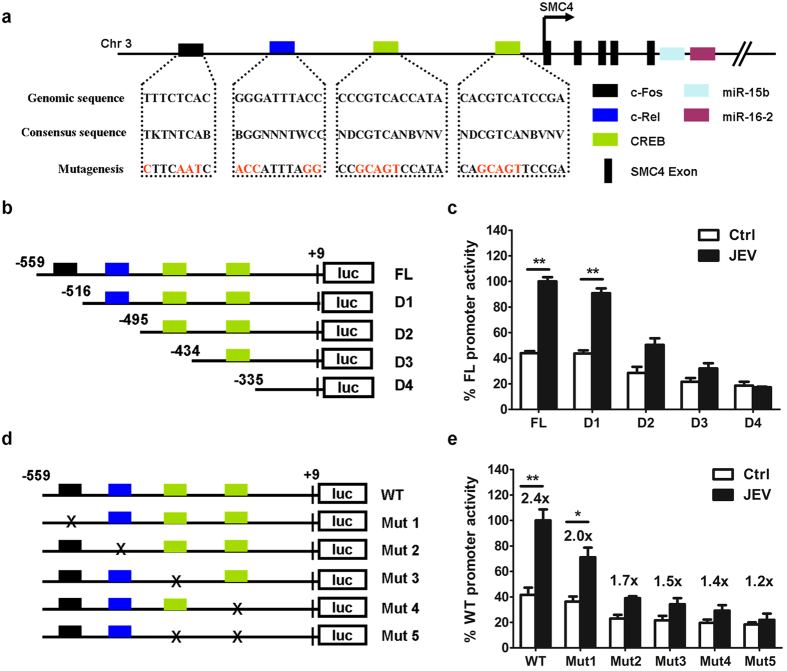
Figure 4. C-Rel and CREB regulate miR-15b promoter activity.
(a) Schematic diagram of miR-15b genomic loci on human chromosomes 3. Putative binding sites of c-Fos, c-Rel, and CREB transcriptional factors (TFs) are shown as boxes. The sequences of mutated sites are shown under the locus diagram. (b) Schematic representation of deletion mutants D1 to D4 in the full-length miR-15b promoter (FL). (c) The promoter reporter constructs (100 ng) described in (b) together with PRL-TK (10 ng) were co-transfected in Hela cells. At 24 h post-transfection, cells were infected with medium or JEV for another 24 h. Samples were collected and analyzed for dual luciferase activity. Results were plotted as firefly luciferase activity (standardized to Renilla luciferase activity) and expressed as percentages of JEV-inducible FL promoter activity (100%). (d) Schematic representation of point mutations (Mut1 to Mut 5) in the wild-type promoter (WT). Mutations, disrupting transcription factor binding, introduced in the promoter construct, which was indicated by x. (e) Hela cells were co-transfected with mutations in (d) and PRL-TK as described in (c). Fold increase after JEV infection relative to basal condition was indicated for each construct. Error bars represent the standard deviation (SD) calculated from results of at least three independent experiments. Statistical analysis was carried out by 2-way ANOVA with subsequent t tests using a Bonferroni post-tests. *p < 0.05; **p < 0.01.

