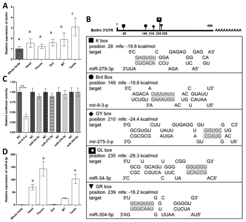
Figure 1. Mitoferrin is most abundantly expressed in testes and targeted by miR-8-3p.
(A) Expression pattern of the B. dorsalis gene mitoferrin in different tissues (whole body, head, thorax, gut, malpighian tube (Mt) and testis) of 12 d old adult males (n = 20). Mitoferrin expression in tissues is relative to whole body expression. The letters above the bars show significant differences (Least Significant Difference in one-way analysis of variance, P < 0.05) in bmfrn expression. The data represent the mean of three independent experiments. Error bars indicate SD. (B) Potential miRNA target sites of miR-279-3p, miR-8-3-p, miR-275-3p, miR-34-3p and miR-304-5pin the 3′-UTR of the mitoferrin as detected by RNAhybrid. Seed sequence of the miRNAs and their putative binding sites in the 3′-UTR are indicated by grey shading. mfe:match free energy. (C) Dual-luciferase assay in HeLa cells co-transfected with psiCHECK-2 -bmfrn 3′-UTR (100 ng) together with negative control miRNA (miR-NC) or miRNA mimics (50 nM) as indicated. Data represent means of three independent experiments, error bars indicate SD. ****P < 0.001, ANOVA with Bonferroni’s Multiple Comparison Test, testing selected pairs (miRNA vs respective miR-NC). (D) Expression pattern of miR-8-3p in different tissues (whole body, head, thorax, gut, malpighian tube (Mt) and testis) of 12 d old adult males (n = 3). Expression in tissues is relative to whole body expression. “a” above the bars indicates significant differences in miR-8-3p expression compared with whole fly homogenate. The data represent the means with SD. N = 3.

