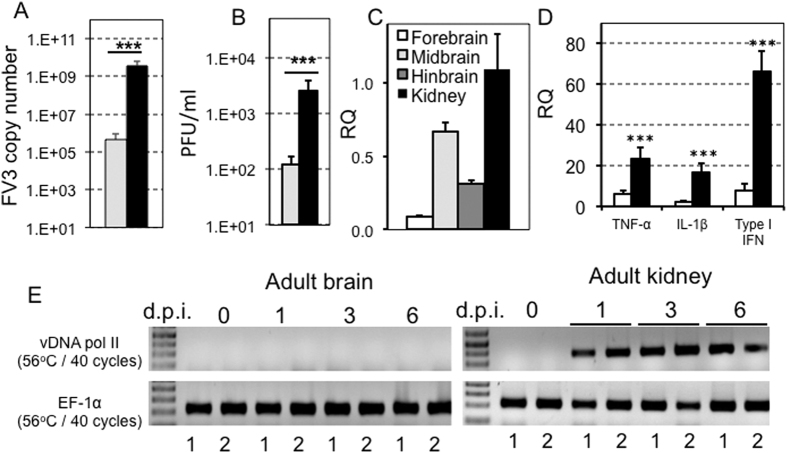
Figure 1. FV3 dissemination to the brain of tadpole but not adult X. laevis.
Outbred pre-metamorphic tadpoles or adults were infected by i.p. injection of 1 × 10⁴ PFU or 1 × 10⁶ PFU of FV3, respectively. (A) FV3 genome copy number of tadpole brains (gray bars) and kidneys (black bars) at 6 d.p.i. (N = 6), determined by absolute qPCR using primers specific for FV3 vDNA Pol II. Results are means ± SE of the FV3 genome copy number per 50 ng of total DNA from 6 animals. ***P < 0.004 significant differences relative to tadpole brains by T-test. (B) Viral loads in tadpole brains (gray bars) and kidneys (black bars) at 6 d.p.i. (N = 6), determined by plaque assay. Results are representative of 3 replicates and displayed as means ± SE of the PFU/mL from 10 animals. ***P < 0.001 significant differences relative to tadpole’s brain by T-test. (C) Viral transcription in tadpole forebrain (white bars), midbrain (clear gray bar), hindbrain (dark gray bar) and kidneys (black bar) at 6 dpi animals, determined by qRT-PCR using primers specific for FV3 vDNA Pol II. P < 0.001 significant differences of each brain section relative to kidney using one-way ANOVA test and Tukey as post hoc test. There was no significant difference among the brain sections. (D) Change in the expression of the pro-inflammatory genes TNF-α and IL-1β as well as the antiviral gene type I IFN, in tadpole brains of 6 days post-FV3 (black bars) or sham-infected (white bars) animals, by qRT-PCR. Primers specific for Xenopus GAPDH (glyceraldehyde-3-phosphate dehydrogenase) were used as an endogenous control, and the expression of these genes were normalized to GAPDH. Results are means ± SE of gene expression from 6 animals. ***P < 0.001 significant differences relative to uninfected tadpole’s brain by T-test. (E) Detection of FV3 infection in two year-old X. laevis adult frog brains and kidneys (2 individuals per group) at 0, 1, 3 and 6 days post-infection. The presence of FV3 was detected in extracted DNA by PCR using primers specific for FV3 vDNA Pol II. EF-1α was use as a housekeeping gene control.

