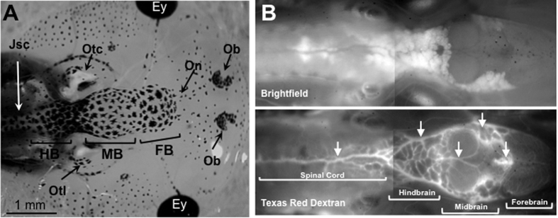
Figure 4. Visualization of tadpole’s brain and blood vasculature.
(A) Dorsal view of a tadpole head at developmental stage 55 under a stereomicroscope at low magnification depicting the forebrain (FB), midbrain (MB), hindbrain (HB) and the junction with the spinal cord (JsC). Other anatomical structures visible are: Ey, eye; Ob, Olfactory bulb; On, olfactory neuron, Otc, Otocyst; Otl, Otolift. (B) Albinos outbred pre-metamorphic tadpoles were injected intracardially with Texas red dextran and 20 min later were anesthetized and observed under a fluorescent microscope with a low (5x) magnification objective. Arrows indicate major blood vessels.

