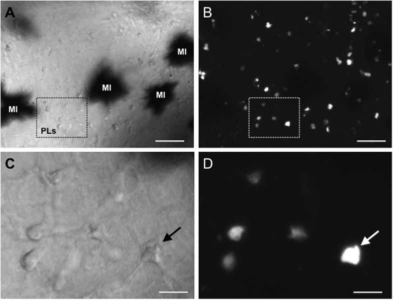
Figure 6. Peritoneal leukocyte transmigration across the BBB in tadpoles.
Whole mount brain preparation from a tadpole at 6 d.p.i adoptively transferred by intracardiac injection of infected PLs labeled with PKH26. The midbrain area was examined under phase contrast (A,C) and fluorescence microscopy (B,D) at low magnification (10x objective) and high magnification (40x objective). The black spot in A are melanophores (MI). The arrow indicates the same cell. White bar =10 µm.

