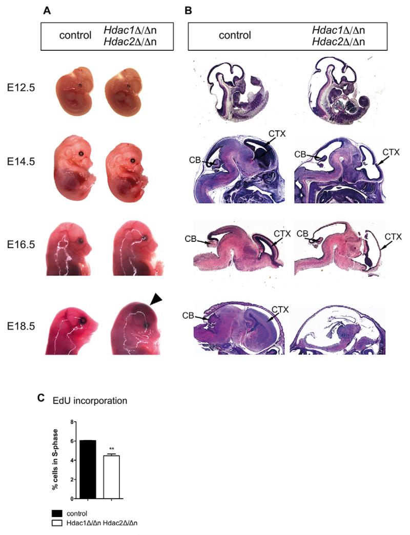
Fig. 3. Combined deletion of Hdac1 and Hdac2 in the nervous system leads to embryonic lethality.
(A) Representative pictures of Hdac1Δ/ΔnHdac2Δ/Δn (right) and wild-type littermate controls (left) at consecutive embryonic time points (E12.5, E14.5, E16.5 and E18.5). The black arrowhead indicates a region affected by brain hemorrhage. (B) Hematoxylin and Eosin stainings of Hdac1Δ/ΔnHdac2Δ/Δn (right) and wild-type littermate control representative paraffin sections (left) at indicated embryonic time points (E12.5, E14.5, E16.5 and E18.5). Cortex and cerebellum are indicated. (C) Quantification of S-phase cells monitored by 5-ethynyl-2′-deoxyuridine (EdU) incorporation and subsequent fluorescence-activated cell sorting (FACS) analysis in E14.5 Hdac1Δ/ΔnHdac2Δ/Δn (white) and control littermate (black) brains. Error bars indicate s.d. (n=3). **P<0.01. CB, cerebellum; CTX, cortex.

