
Figure 4.
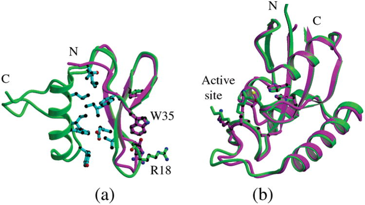
Domain structure comparison of CaEss1 with human Pin1. (a) Superposition of the WW domain of Pin1 (magenta) onto the WW domain and linker of CaEss1 (green) showing the overall similarities of the folds of the three-stranded β-sheet domains. The residues that form a hydrophobic core between the WW domain and the linker are shown in cyan. (b) Superposition of the PPIase domains of CaEss1 (green) and Pin1 (magenta).