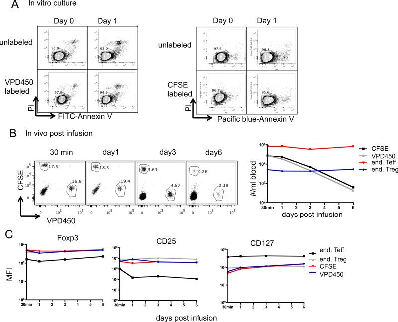
Figure 2. CFSE / VPD450 labeling does not affect the survival of expanded Treg in vitro or in vivo.
Expanded Treg (round 3) were labeled with CFSE or VPD450. (A) They were then cultured in the presence of 300 U/ml of IL-2 for an additional day, and apoptosis analysed by staining with Annexin V and 7-AAD. Unlabeled cells were used as controls. Data are representative of 3 independent experiments. (B, C) Expanded non-auto Treg were labeled with CFSE or VPD450, equal numbers of labeled cells mixed together and infused into an IS monkey (CM118). At the indicated time points, 1ml blood was tested for the presence of infused cells and end Teff and end Treg, as described in Figure 2. (B) dot plots and plot of #/ml blood vs. time post-infusion are shown. (C) MFI of Foxp3, CD25, CD127 in/on infused CFSE-labeled or VPD450-labeled exogenous Treg were compared with those in/on end Teff and end Treg at the indicated time points.

