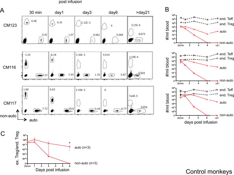
Figure 3. Persistence of ex vivo-expanded Treg in peripheral blood of control monkeys.
Auto- or non-auto-Treg were labeled with either CFSE or VPD450, respectively, then infused i.v. into healthy control monkeys. At the days indicated post-Treg infusion, 1ml blood was tested for the presence of the infused cells and endogenous (end) Teff and Treg by flow cytometry. Counting beads were added before running flow analysis. Cell numbers per ml blood were calculated as the number of cell events/number of bead events × total number of beads added. Duplicate samples were tested at each time-point. (A) Flow cytometric profiles of CD4-gated PBMC from 3 monkeys showing the percentages of infused, labeled auto-and non-auto-Treg. Endogenous (unlabeled) CD4+ T cells are also evident (lower left quadrant). (B) Kinetics of the number of infused Treg (auto- and non-auto-Treg) and endogenous (end) Teff and Treg numbers in the peripheral blood after labeled Treg infusion in the 3 control monkeys. (C) Kinetics of the ratio of infused (ex) Treg to endogenous (end) Treg in the peripheral blood after labeled Treg infusion in these monkeys (n=3). *, The ratio of auto-Treg/end Treg was significantly greater than that of non-auto-Treg/end Treg, p<0.05.

