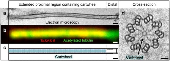
Fig. 2.

Exceptionally long basal body in Trichonympha. a Electron micrograph of isolated Trichonympha sp. basal body. b Immunofluorescence of isolated Trichonympha sp. basal body revealing TaSAS-6 localization (red, yellow in overlay) along the basal body/flagellum complex stained with acetylated tubulin (green). c Schematic representation of the exceptionally long basal body of Trichonympha, with the cartwheel-bearing region highlighted. d Cross section of Trichonympha basal body; arrow points to cartwheel structure, with central hub and nice radial spokes connecting with peripheral microtubules. Scale bar in (a, b) 250 nm, in (d) 50 nm
