Figure 6.
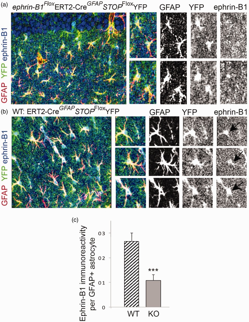
Specific ablation of ephrin-B1 from adult hippocampal astrocytes but not CA1 neurons in vivo. (a, b) Confocal images show YFP (green), GFAP (red), and ephrin-B1 (blue) in the SR area of CA1 hippocampus of ephrin-B1Flox/yGFAP-ERT2Cre/+STOPFloxYFP (KO, A) and control GFAP-ERT2Cre/+STOPFloxYFP (WT, B) mice. GFAP and ephrin-B1 levels were detected by immunostaining. YFP-positive WT astrocytes (black arrow, b) but not YFP-positive KO astrocytes (a) express ephrin-B1. Note that the ephrin-B1 deletion is specific to astrocytes and CA1 neurons express ephrin-B1 in both WT and KO mice. (c) Graph shows mean integrated intensity of ephrin-B1 immunoreactivity per GFAP-positive astrocyte in WT (n = 552 cells, 9 images, 3 mice) and KO (n = 520 cells, 9 images, 3 mice) groups (mean ± SEM; n = 3 mice). Statistical analysis was performed using paired Student’s t test (*p < .05, **p < .01).

