Figure 7.
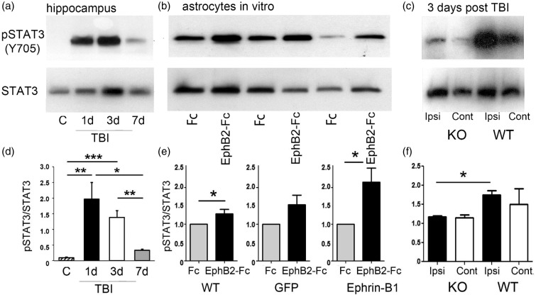
STAT3 phosphorylation in astrocytes is regulated by ephrin-B1. (a) Western blot analysis of pSTAT3 and STAT3 in the hippocampus of control, 1, 3, or 7 dpi. (b) Western blot analysis of pSTAT3 and STAT3 in the primary cultures of untransfected astrocytes (WT) and astrocytes overexpressing GFP (GFP) or ephrin-B1 (ephrin-B1) following the treatment with control Fc or EphB2-Fc for 15 min. (c) Western blot analysis of pSTAT3 and STAT3 in the ipsilateral (Ipsi) or contralateral (Cont) hippocampus of ephrin-B1 KO or WT at 3 dpi. The blots were first probed against pSTAT3 and then reprobed for total STAT3. (d–f) Graphs show pSTAT3/STAT3 (mean ± SEM; n = 3–4 cultures for E, Student’s t test, *p < .05; n = 3–4 mice for d and f, one-way ANOVA followed by Tukey’s post hoc analysis, *p < .05, **p < .01, ***p < .001). The levels of pSTAT3 and total STAT3 in EphB2-Fc-treated cultures were normalized to Fc-treated cultures (e).

