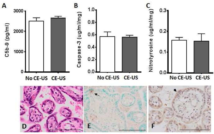
Figure 3. Japanese macaque placental tissue integrity with and without contrast-enhanced ultrasound exposure.

(A) Complement C5b-9 expression in maternal serum obtained at the time of G130 cesarean section delivery, (B) Caspase 3 and (C) Nitrotyrosine protein expression in placental homogenate from control animals who were not exposed to CE-US (white bar) and CE-US exposed (grey bar) animals. Data are mean±SEM for n=6 animals/group. Representative placental tissue sections from a control animal with microbubble exposure during ultrasound stained for (D) Hematoxylin and Eosin (E) TUNEL, where the arrow highlights an apoptosis-positive cell and (F) HSP70, where the arrowhead indicates positive staining on the syncytiotrophoblast. Scale bar indicates 100μm.
