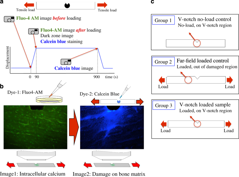
Figure 4.
(a) Loading history and experimental design for applying stains for sequential imaging of intracellular calcium fluorescence and matrix damage on the same sample. (b) Representative images obtained from dual staining. MC3T3-E1 cells were stained with Fluo4-AM for evaluating intracellular calcium signaling, and damage in bone matrix was stained with calcein blue. (c) Study groups.

