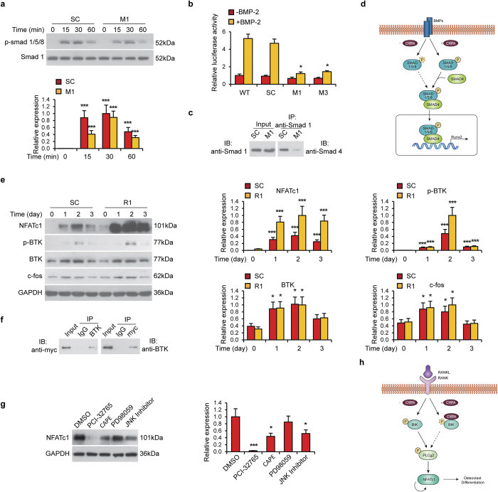
Figure 4. CypA affects mouse osteoblast and osteoclast via different signaling pathways.
(a–d) CypA regulation of BMP-2 induced Smad phosphorylation in osteoblastic cells. (a) SC and M1 MC3T3-E1 osteoblastic cells were treated with BMP-2. Cells were harvested for western blot of p-Smad1/5/8 expression at various timepoints. Smad1 was used as a loading control. (b) Id1 Luciferase activity of WT, SC, M1 and M2 cells in the presence of BMP2. Data represented as mean and SEM from three independent experiments. *P < 0.05 by one way ANOVA analysis, compared to WT cells in the same group. (c) Co-immunoprecipitation between Smad1 and Smad4 in SC and M1 MC3T3-E1 osteoblastic cells. (d) Schematic diagram of CypA in osteoblastic differentiation. E-H CypA regulation of downstream signaling in osteoclastic cells. (e) p-BTK, BTK, c-fos and NFATc1 levels in SC and R1 RAW263.7 osteoclastic cells during osteoclastic induction at various timepoints as assessed by western blot. GAPDH was used as a loading control. (f) Myc-tag CypA plasmid was transfected into RAW264.7 cells for two days, follow by co-immunoprecipitation assay between myc-CypA and BTK. (g) NFATc1 levels in differentiated RAW264.7 cells (R1 clone) treated with PCI-32765, CAPE, PD98059 or JNK inhibitor for two days. GAPDH was used as a loading control. (h) Schematic diagram of CypA in osteoclastic differentiation.

