Fig. 1.

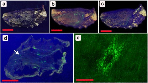
Fluorescent signals from the Dll-GFP fusion protein expressed in pupal wings after baculovirus-mediated gene transfer. a A non-treated pupa under the blue illuminator. Scale bar, 5 mm (also applicable to b-d). b, c The Dll-gfp baculovirus-treated pupae under the blue illuminator. Green areas signify GFP fluorescent signals. d An isolated forewing that exhibits many patches of GFP fluorescence. An arrow indicates a single GFP-positive patch that is enlarged in e. e A GFP-positive patch. Numerous small green dots are epithelial cells expressing Dll-GFP. Scale bar, 200 μm
