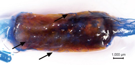
Figure 2.

Tissue-engineered nerve after clearing with glycerin.
The proximal end is on the right, and the distal end on the left. The nerve stumps at the ends of the tissue-engineered nerves and connective tissues surrounding the tissue-engineered nerves were transparent. The chitosan nerve conduits appeared semitransparent. Both the small vessels in the nerve stumps and the microvessels growing into the tissue-engineered nerves were filled with blue contrast agent (arrows) and are clearly visible. The microvessels growing into the tissue-engineered nerves from the surrounding tissues are indicated with arrows.
