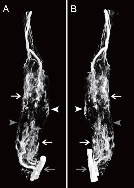
Figure 3.

Three-dimensional reconstructions of blood vessels in tissue-engineered nerve.
(A, B) Views from two different angles of a three-dimensional reconstruction. The proximal end is at the top, and the distal end at the bottom. The blood vessels of the popliteal fossa are indicated by the gray arrow. A large number of microvessels and capillaries were relatively well visualized. Microvessels growing into the tissue-engineered nerves are indicated with white arrows. New blood vessels grew into the tissue-engineered nerves from three main directions: the proximal end, the distal end, and the middle. The microvessels growing in from the middle part are indicated with white arrowheads. The microvessels connecting the proximal and distal ends are indicated with gray arrowheads.
