Figure 2.

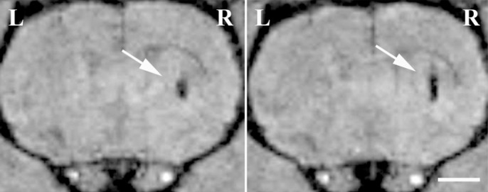
MRI of hVM-MNP labeled cells transplanted into rat brain.
Two consecutive in vivo magnetic resonance images of hVM cell suspensions (3 × 10⁵ cells) transplanted into the left (hVM cells, L) and right (hVM-MNP labeled cells, R) striata. MRI was performed eight weeks after cell transplantation. hVM-MNP labeled cells can be easily detected in coronal T2*-weighted images as dark hypointense signals in the area where the cells have been injected (arrows). No MRI signal was detected when unlabeled hVM cells were transplanted (left striatum, L). Scale bar: 2 mm. hVM: Human ventral midbrain; MNP: magnetic nanoparticle; MRI: magnetic resonance imaging; R: right stratum.
