
Figure 1.
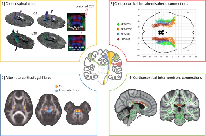
Networks of interest in structural connectivity analyses after stroke. This figure is to illustrate how structural imaging can be used to study stroke‐related changes in structural connectivity in different networks. The CST originating from the primary motor cortex has been studied by numerous studies (represented in yellow): For example, diffusion tensor imaging has been used to reconstruct the CST in a 47‐year‐old man 3 (d3) and 30 days (d30) after left‐sided stroke. Reduction in fractional anisotropy in the lesioned left CST at pons level at d30 (FA, rFA values compared to the contralateral side) was regarded as Wallerian degeneration (1, adapted with permission).45 Recent imaging data have suggested that not only the CST but also alternate motor fibers might contribute to motor functioning and recovery after stroke (represented in blue): For instance, tractography was used to reconstruct such alternate fibers (in blue), probably paralleling the cortico‐rubro‐spinal system, in addition to the CST (2, adapted with permission).38 Intrahemispheric corticocortical connections (in red) have been addressed by more recent analyses, for instance, between frontal and parietal motor areas. It could be shown that aside from the CST, also parietofrontal structural connectivity relates to residual motor function after stroke. M1 primary motor cortex, PMv ventral premotor cortex, aIPS anterior/cIPS caudal intraparietal sulcus (3, adapted with permission from Robert Schulz et al. Parieto frontal motor pathways and their association with motor function after stroke. Brain (2015) 138 (7): 1949–1960. (Fig. 1.) By permission of Oxford University Press on behalf of The Guarantors of Brain. This image is not covered by the terms of the Creative Commons/Open Access license of this publication. For permission to reuse, please contact the rights holder).22 Ultimately, also structural connections between both hemispheres have been evaluated in regard of their contribution for motor functioning and recovery after stroke. For example, regional FA values along the corpus callosum were related to residual motor function in the chronic stage of recovery (4, adapted with permission).72 Notably, applications of large‐scale network analyses were not included in this synopsis for the sake of clarity. CST, corticospinal tract; FA, fractional anisotropy; rFA, relative FA.