Fig. 5.
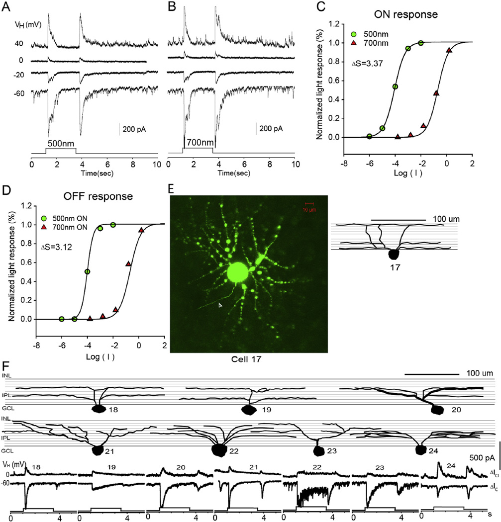
Medium-dendritic-field ON–OFF RGC. The current responses evoked by 2.5 s 500 nm light (A) and 700 nm light (B) of unattenuated 0 log unit intensity at various holding potentials; (C and D) the stimulus intensity–response relations for the ON and OFF responses; (E) the stacked confocal fluorescent image in the flat-mounted retina and the open triangle points to the axon; (F) sketches of medium-dendritic-field ON–OFF RGCs on a schematic background of the inner plexiform layer, and corresponding LePSCs evoked by 2.5 s 500 nm light at holding potentials near ECl (ΔIC) and near EC (ΔICl) for each cell. INL: inner nuclear layer; IPL: inner plexiform layer; RGC: retinal ganglion cell layer.

