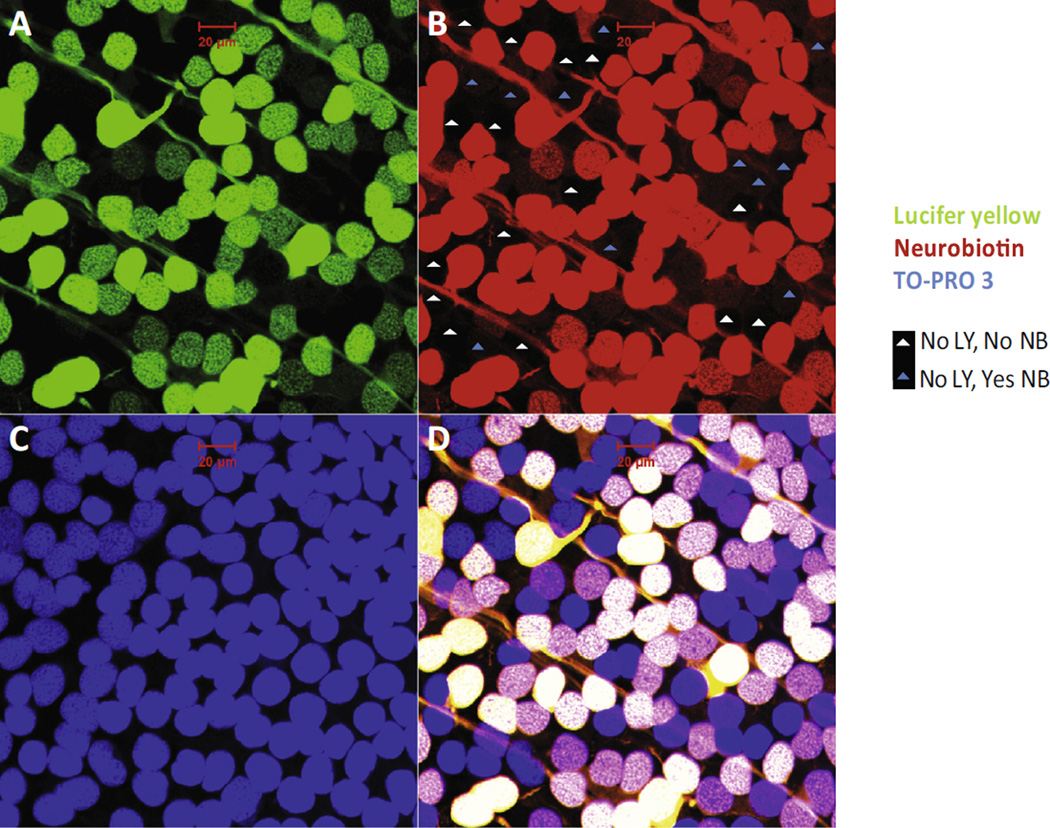
Fig. 9.
Displaced amarine cells in the RGC layer. In the RGC layer, retrograde–identified RGCs with both LY and NB labeling constituted 3/4 of the total neurons; the remaining 1/4 of neurons with no LY signal were displaced amacrine cells which are either coupled to RGCs (with NB signal) or not coupled to RGCs (without NB signal). (A) LY labeling, green; (B) NB labeling, red; (C) TO-PRO-3 labeling, blue; (D) combined. Scale bar: 20 µm. (For interpretation of the references to color in this figure legend, the reader is referred to the web version of this article.)

