Figure 3.

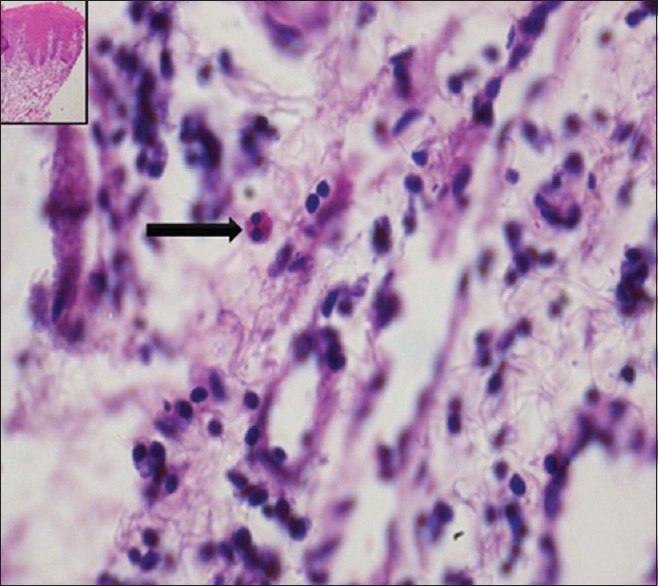
The section shows tissue eosinophil in a case of moderate epithelial dysplasia (H&E stain, x400). Inset: Scanner view of the lesion (H&E stain, x40)

The section shows tissue eosinophil in a case of moderate epithelial dysplasia (H&E stain, x400). Inset: Scanner view of the lesion (H&E stain, x40)