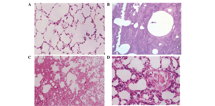
Figure 2.
Hematoxylin and eosin stained sections of lung tissue harvested from various groups. (A) Group A: Normal pulmonary tissue morphology and no pulmonary edema detected (magnification, ×100). (B) Group B (magnification, ×40) and (C) group C (magnification, ×10): Pulmonary congestion and edema were detected, with a protein rich liquid saturating the pulmonary interstitium, alveoli and bronchioles. (D) Group D: Pulmonary interstitial edema and thickening were detected, as well as inflammatory cell infiltration, while the alveolar cavity was filled with exudation (magnification, ×20).

