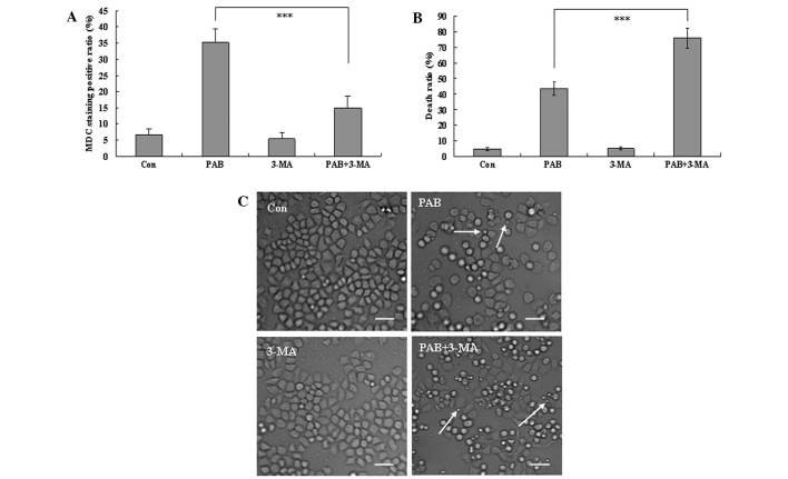
Figure 3.
3-MA inhibits autophagy and promotes cell death in PAB-treated MCF-7 cells. (A) The positive ratio of MDC staining (autophagic ratio) was analyzed quantitatively by flow cytometry. PAB increased MDC staining which was inhibited by 3-MA. (B) 1×10⁴ MCF-7 cells/well were incubated with 4 µM PAB or 3 mM 3-MA for 36 h and growth inhibition was evaluated using the MTT assay. These findings indicated that 3-MA increased the efficacy of PAB in inducing cell death. Error bars = mean ± standard deviation, ***P<0.001. (C) 3-MA in addition to PAB promoted cell death following treatment with 4 µM PAB for 36 h. Arrows indicate apoptotic bodies. Scale bar, 25 µm. All experiments were performed in triplicate. MDC, monodansylcadaverine; Con, control; PAB, pseudolaric acid B; 3-MA, 3-methyladenine.

