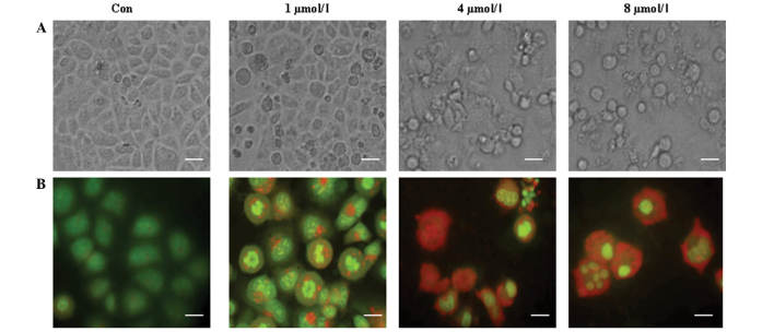
Figure 5.
PAB increased autophagy and apoptosis in a dose-dependent manner. (A) Cell morphology was visualized by phase contrast microscope after 0, 1, 4 and 8 µM PAB treatment for 36 h, revealing that PAB more effectively induced cell death with increasing doses (scale bar, 20 µm). (B) Acridine orange staining of autophagic vacuoles was visualized in MCF-7 cells treated with 0, 1, 4 and 8 µM PAB for 36 h, indicating that PAB increased the number of autophagic vacuoles with increasing doses (scale bar, 10 µm). The experiments were performed in triplicate. Con, control.

