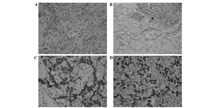
Figure 2.
Histopathological analysis of bronchial biopsy specimens. (A) Hematoxylin and eosin staining revealing (A) poorly differentiated squamous cell carcinoma (magnification, ×200) and (B) intercellular bridges (indicated by the black arrow) of squamous cell carcinoma (magnification, ×200). (C) Immunohistochemical staining revealing positivity for (C) CK5/6 (magnification, ×200) and (D) p40 (magnification, ×200) within the areas of carcinoma nests.

