Figure 4.
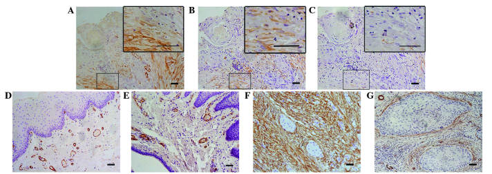
Immunohistochemical staining of CAFs. CAFs were (A) α-SMA and (B) vimentin positive, but (C) desmin negative. (D and E) Negative staining of α-SMA in the epithelial dysplasia and paired tumor-adjacent non-neoplastic tongue epithelium samples. (F) Typical ‘network’ pattern formed when CAFs are particularly abundant and occupy almost the entire tumor stroma. (G) ‘Spindle’ pattern, in which CAFs are arranged in 1–3 rows in a regular order in the periphery of the neoplastic islands. CAFs, cancer-associated fibroblasts; SMA, smooth muscle actin.

