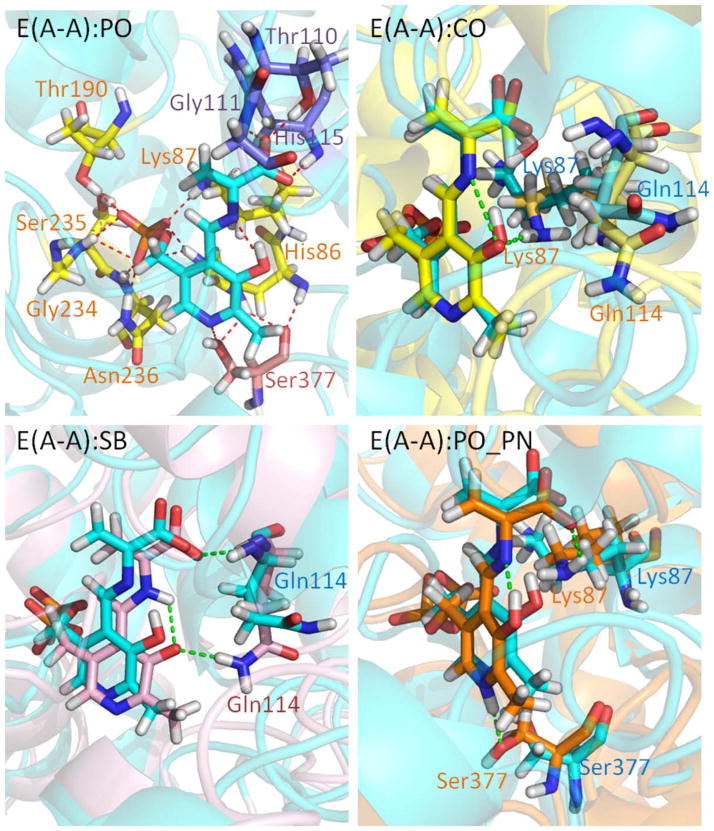
Figure 3.
Protonation states of E(A-A). (PO) Presentation of E(A-A):PO binding to TRPS. The residues interacting with the phosphoryl group (PG), PN, and carboxylate oxygens (COs) of E(A-A):PO are shown in yellow, pink, and purple, respectively. (CO) The alignment of E(A-A):CO (yellow) and E(A-A):PO (cyan). Residues Lys87 and Gln114 are shown. The H-bond between Lys87 and the PO of E(A-A):CO is shown in green. (SB) The alignment of E(A-A):SB (pink) and E(A-A):PO (cyan). The Gln114 side chain forming H-bonds with the PO of E(A-A):SB is shown as a green dashed line. (PO_PN) The alignment of E(A-A):PO_PN (orange) and E(A-A):PO (cyan) is depicted. The Lys87 and Ser377 residues can form H-bonds with E(A-A):PO_PN. All snapshots are from 50-ns MD simulation.

