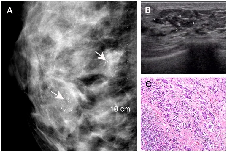
Figure 3.
A 42-year-old breast cancer patient with residual disease of the right breast following lumpectomy. (A) Mammography revealed dense breast tissue with multiple clustered microcalcifications (arrows). (B) Ultrasonography revealed an indistinct hypoechoic area with internal hyperechoic foci (asterisks), corresponding to the mammographically visualized microcalcifications. (C) Histological diagnosis of residual invasive ductal carcinoma (haematoxylin and eosin staining; magnification, ×10).

