Fig. 5.
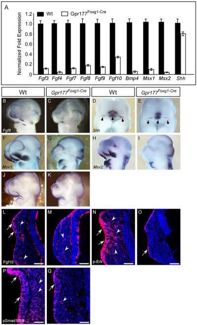
FGF, BMP, and SHH signaling pathways are altered in the facial prominence of Gpr177Foxg1-Cre embryos. A: Quantitative RT-PCR analysis of gene expression in the facial prominences of E10.25 embryos. Expression of Fgf3, Fgf4 Fgf7, Fgf8, Fgf9, Fgf10, Bmp4, Msx1, and Msx2 is significantly down-regulated in Gpr177Foxg1-Cre embryos. By contrast, expression of Shh is mildly decreased. B–K: Whole-mount in situ analysis shows that expression of Fgf8, Shh, Msx1, Msx2, and Bmp4, in the facial prominences of E10.5 Gpr177Foxg1-Cre embryos as presented in sagittal views. Note that the expression of Fgf8, Msx1, and Msx2, is abolished completely in lnp, mnp, and mxp of Gpr177 mutant embryos. D,E: Frontal facial views of the wild-type and mutant embryos show that in Gpr177Foxg1-Cre embryos, the expression of Shh is retained in the FEZ (Arrowheads); however, its expression region in telencephalon and diencephalon becomes smaller (Arrows). Immunofluorescence analysis on sections of E10.25 facial primordia for Fgf10 (L,M), p-Erk (N,O), and pSmad1/5/8 (P,Q). Expression of Fgf10 protein is reduced in both the epithelium (arrows) and the mesenchyme (arrowheads) of embryos lacking epithelial Gpr177. Note that, in the facial primordia of wild-type embryos, distribution of Fgf10 proteins is concentrated at the sub-epithelial mesenchyme where the epithelium recesses (L). However, Fgf10 proteins are scattered in the facial mesenchyme of Gpr177Foxg1-Cre embryos (M). Phosphorylation of Erk of Gpr177Foxg1-Cre embryos is significantly reduced in the epithelium and diminished in the mesenchyme (O vs. wild-type N). Phosphorylation of Smad1/5/8 of Gpr177Foxg1-Cre embryos is reduced in the olfactory epithelium and diminished in the mesenchyme (Q vs. P). Arrows and arrowheads point out positive staining in the epithelium and the mesenchyme, respectively. Scale bars ¼ 100 mm. FEZ, frontonasal ectodermal zone.

