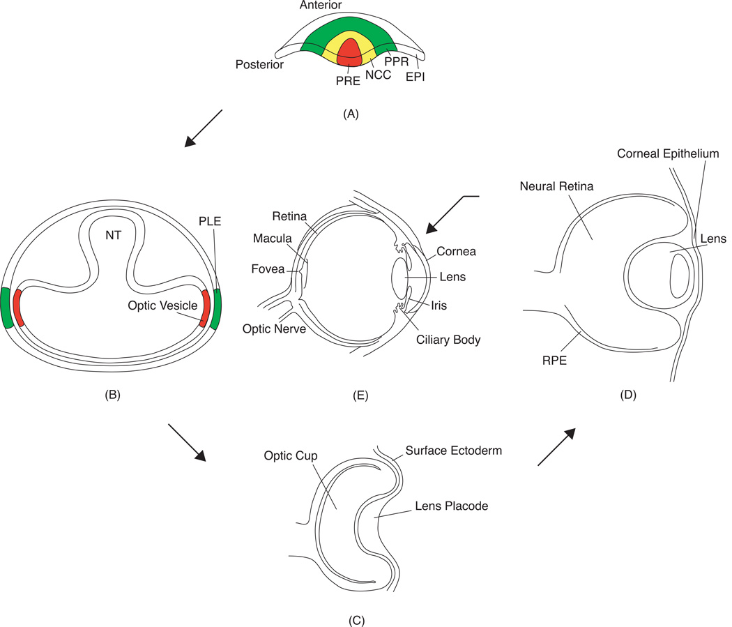
Fig. 1.
Vertebrate ocular development. (A) In late gastrulation, the anterior neural plate contains the presumptive retinal ectoderm (PRE) surrounded by neural crest cells (NCC), pre-placodal region (PRR), and the epidermis (EPI). (B) As the neural tube (NT) closes, the diencephalon bilaterally develops into the optic vesicles, which contact the presumptive lens ectoderm (PLE) on either side. (C) Co-ordinate signaling leads to induction of the PLE to form the lens placode, while the optic vesicle invaginates to form the optic cup. (D) The lens placode invaginates and forms the lens pit that detaches from the surface ectoderm to form the lens vesicle. At this stage the optic cup starts to differentiate into the neural retina and the retinal pigment epithelium (RPE). (E) The adult vertebrate eye thus contains multiple tissue compartments. It is important to note that in fish and frogs, the lens placode does not form a hollow lens vesicle, but instead develops into a solid aggregate of cells.

