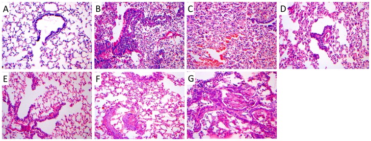
Figure 2.
Pathological changes in lung tissues obtained from tree shrews observed under a light microscope, following hematoxylin and eosin staining; magnification, ×200. (A) Tree shrews normal lung epithelial structure in the solvent control group in the 11th week. (B) Infiltration of inflammatory cells in lung tissue in the 3rd week (animal in the experimental group). (C) Alveolar hemorrhage, bronchial epithelial and other pathological changes of tree shrews in the experimental group in the 3rd week. (D) Mild atypical hyperplasia of bronchial epithelial in a tree shrew from the experimental group in the 5th week. (E) Bronchial epithelial moderate dysplasia change in tree shrews from the experimental group in the 7th week. (F) Bronchial epithelial dysplasia change observed in tree shrews from the experimental group in the 9th week. (G) Bronchial epithelial carcinoma in situ in an animal from our experimental group in the 11th week.

