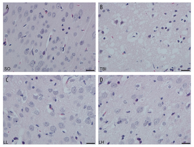
Figure 1.
The HE staining of brain tissue in different groups after 24 h. (A) SO group, (B) TBI group, (C) LL group, (D) LH group. There was no significant pathological lesion in brain tissue slice of SO group. The swollen cells were successively alleviated in TBI, LL, and LH groups. Scale bar: 100 μm (×400).

