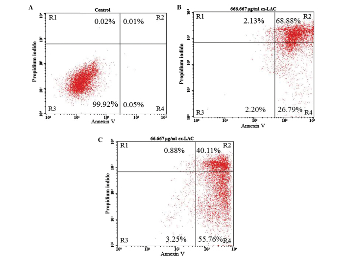
Figure 3.
Cytotoxic effect of ex-LAC on Jurkat cells. (A) Control cells. (B) Cells treated with 666.667 µg/ml ex-LAC. (C) Cells treated with 66.667 µg/ml ex-LAC. Cytotoxicity was assessed by staining the cells with Annexin V and PI prior to being subjected to flow cytometry analysis. In each graph, the lower left quadrant (R4) indicates viable cells (Annexin V−PI−); the upper left quadrant (R2) represents necrotic cells (Annexin V−PI+); the lower right quadrant (R5) indicates early apoptotic cells (Annexin V+PI−); and the upper right quadrant (R3) represents late apoptotic cells (Annexin V+PI+). ex-Lac, extracellular laccase; PI, propidium iodide.

