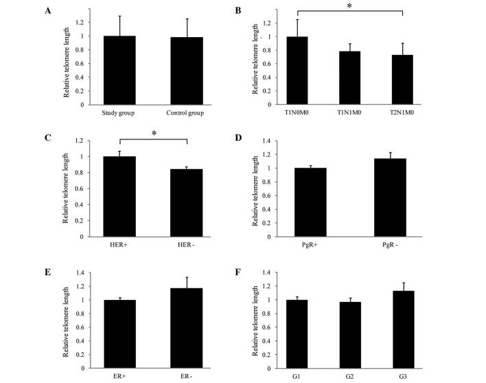
Figure 1.
Quantitative analysis of telomere length in peripheral leukocytes from patients with breast cancer (study group) and control subjects, and the correlation with clinical parameters. Relative telomere length was assessed according to protocol previously described by Cawthon (with modifications) (12) using quantitative polymerase chain reaction. Telomere length was presented relative to a single copy gene albumin. Relative length of telomeres, comparing the (A) study and control groups; (B) tumor stage (according to the TNM classification as follows: T1N0M0, small primary tumor and no lymph node involvement or distant metastases; T1N1M0, small primary tumor, sentinel node metastasis with no distant metastases; T2N1M0, larger primary tumor, sentinel node metastasis with no distant metastases); (C) HER status; (D) PgR status; (E) ER status; and (F) tumor grade. *P<0.05, comparison shown by brackets. T, tumor; N, node; M, metastasis; HER2, human epidermal growth factor receptor 2; PgR, progesterone receptor; ER, estrogen receptor; G1/2/3, grades 1/2/3.

