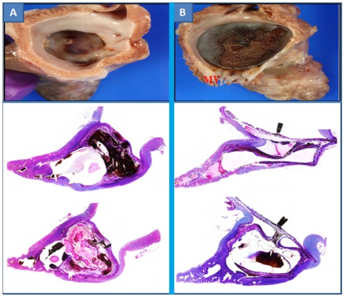
Fig. (3).
Gross and Microscopy images of WM and ACP in a canine model at 28 days. The WM device (A) showed the central area of the device covered with a layer of neo-endocardial tissue with tight device apposition to the native LAA wall. The ACP device (B) showed an area of bare flange mesh wires near the inferior edge of the disk; also there was incomplete coverage of the end-screw hub [28].

