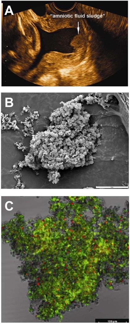
Figure 10.
Microbial biofilms in the amniotic cavity. (A) Two-dimensional transvaginal ultrasound image showing the presence of “amniotic fluid sludge”. (B) Scanning electron micrograph of a floc of “amniotic fluid sludge” showing the bacterial cells and the exopolymeric matrix material which constitute a biofilm. In the center of the image, cocci are resolved amongst a fibrous mass of matrix material. (C) Confocal laser scanning microscopy displays bacteria (red dots), matrix material (green), and some unstained material which is likely to represent host components trapped by the biofilm. The bar represents 100 microns. Bacteria (red dots) are stained with the EUB338-Cy3probe which reacts with the 16S rRNA (component of bacteria). The matrix material has been stained with wheat germ agglutinin, which reacts with the N-acetylglucosamine of the component of the matrix material that forms the structural framework of the biofilm. Modified from Figure 1,3 and 4 Romero R, Schaudinn C, Kusanovic JP, Gorur A, Gotsch F, Webster P, Nhan-Chang CL, Erez O, Kim CJ, Espinoza J, Gonçalves LF, Vaisbuch E, Mazaki-Tovi S, Hassan SS, Costerton JW., Am J Obstet Gynecol. 2008 Jan;198(1):135.e1-5.

