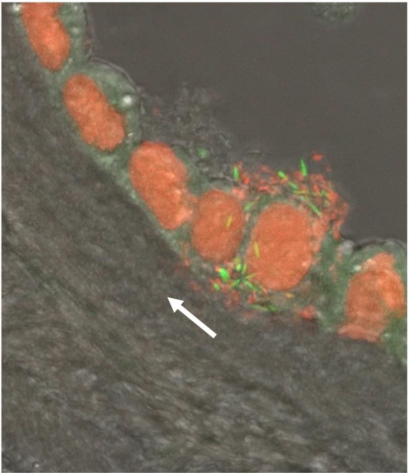
Figure 9.
A cluster of bacteria in amniotic fluid and bacterial invasion of amniotic epithelial cells demonstrated by fluorescent staining. Live bacteria were stained with SYTO 9 (green fluorescence), and dead bacteria were stained with propidium iodide (red fluorescence). Note the lack of bacteria in the chorioamniotic connective tissue indicating bacterial propagation from the amniotic cavity (white arrow). Modified from Figure 3C Kim MJ, Romero R, Gervasi MT, Kim JS, Yoo W, Lee DC, Mittal P, Erez O, Kusanovic JP, Hassan SS, Kim CJ. Lab Invest. 2009 Aug;89(8):924-36.

