Figure-4.

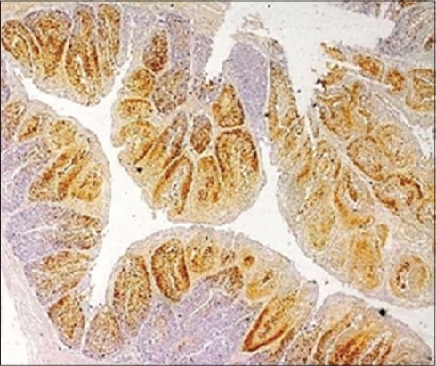
The affected bursal section showing infectious bursal disease viral antigens in the lesion sites identified in bursal follicles (Immunohistochemistry, ×4).

The affected bursal section showing infectious bursal disease viral antigens in the lesion sites identified in bursal follicles (Immunohistochemistry, ×4).