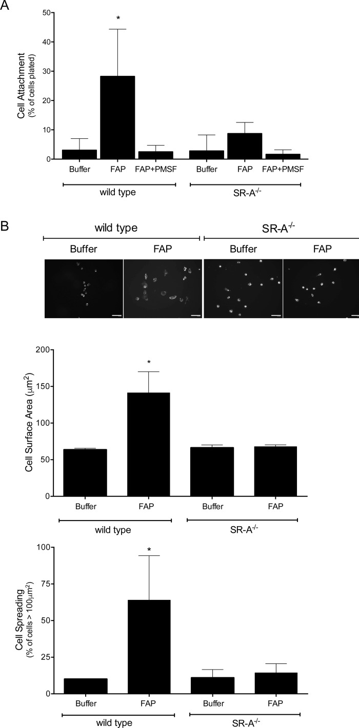
Fig 5. SR-A mediates macrophage adhesion to FAP-modified type I collagen.
(A) MPMs isolated from wild-type (SR-A+/+) or SR-A-/- mice were adhered to type I collagen pretreated with buffer, FAP, or PMSF-inhibited FAP. Non-adherent cells were removed by washing, and the number of attached cells quantified and expressed as a percentage of total cells plated. Shown are the means ± SD of 4 experiments. Results were compared by one-way ANOVA with Tukey’s post-hoc test. (B) MPMs were adhered to type I collagen pretreated with buffer or FAP. Non-adhered cells were removed by washing, and attached cells were fixed and stained with fluorescent phalloidin and DAPI. Representative images were digitally captured and the surface area of cells quantified. Scale bars = 30 μm. Shown are the means ± SD of cell surface area and percentage of cells displaying a surface area > 100 μm2 from 3 experiments. Results were compared by t-test. * indicates significant (p<0.05) difference from wild-type macrophages plated on buffer treated collagen.

