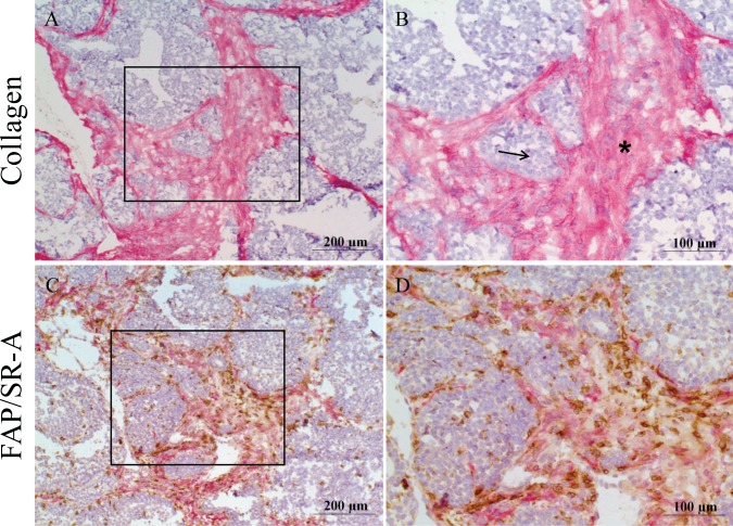
Fig 6. SR-A expressing macrophages localize in the tumor stroma.
Consecutive frozen tumor sections were prepared from MMTV-PyMT mice and immunostained using A-B) a collagen antibody to detect stromal regions (red stain); C-D) a dual-stain technique with a FAP antibody to detect fibroblasts (red stain) and a SR-A antibody to visualize SR-A-positive macrophages (brown stain). Tissues were counterstained with hematoxylin and images digitally captured. B and D represent high power magnification (40x) of inset regions from A and C respectively. Arrows indicate acini of epithelial cells and asterisks indicate collagen-rich stroma.

