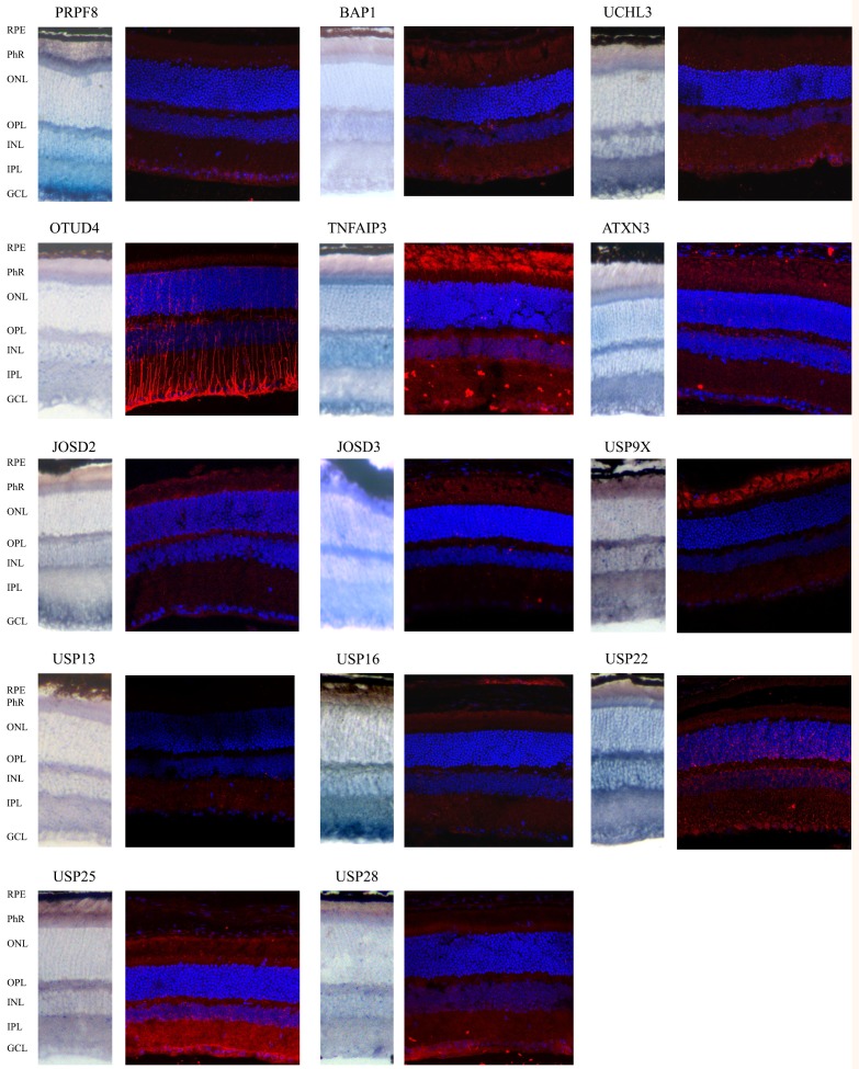
Fig 3. Comparison of mRNA and protein immunodetection of selected DUBs in mouse retinal cryosections.
Most analyzed genes render a consistent expression pattern when comparing mRNA and protein localization in the wild type mouse retina. The merge immunohistochemistry show DUBs immunodetected in red, and nuclei counter-staining with DAPI (in blue). Details in S3 Fig. RPE- Retinal pigmented epithelium; Phr- Photoreceptor cell layer; ONL- Outer nuclear layer; OPL. Outer plexiform layer; INL- Inner nuclear layer, IPL- Inner plexiform layer; GCL- Ganglion cell layer.

