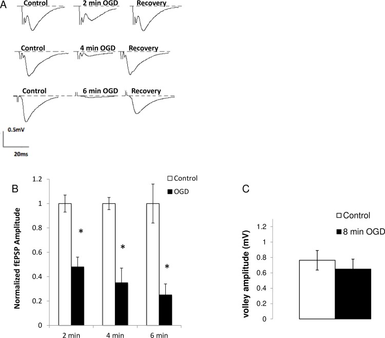
Fig 1. Ischemia depresses the amplitude of evoked synaptic transmission without changes in the presynaptic volley amplitude.
(A). fEPSPs were recorded from the stratum radiatum in the CA1 region while the Schaffer collaterals were stimulated every 15 s. fEPEP amplitude decreased reversibly in a time-dependent manner after 2 min (n = 7), 4 min (n = 5), and 6 min (n = 5) of OGD. (B) fEPSP amplitudes produced by 2 min, 4 min, 6 min of in vitro ischemia relative to controls. (C) Fiber volley amplitude after 8 min of OGD (n = 6, p>0.05) Average plotted as mean ± SE. * p < 0.001: paired student t-test. OGD: Oxygen-glucose deprivation, fEPSP: field excitatory postsynaptic potentials.

