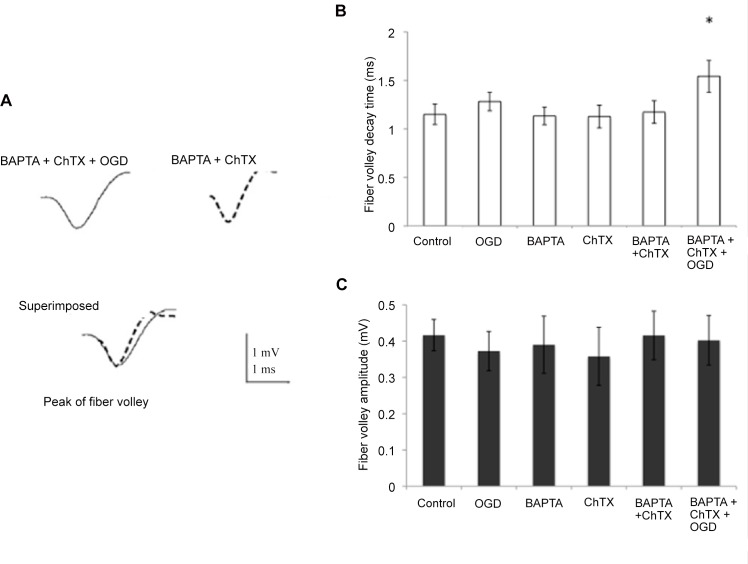
Fig 8. Decay time in the presynaptic component action potential (fiber volley) was lengthened in the presence of OGD and chelator + ChTX.
(A) Fiber volley recorded in the presence of BAPTA-AM + ChTX + OGD (solid line) and in presence of BAPTA-AM + ChTX only (dotted line). Fiber volleys were aligned by their negative peaks and superimposed to compare their amplitude and decay time. (B) Decay time of the fiber volley was increased in BAPTA-AM + ChTX + OGD condition versus BAPTA-AM + ChTX alone. It remained unchanged in control, OGD, BAPTA-AM and ChTX condition. (C) Amplitude of the fiber volley was unchanged in the presence of BAPTA-AM + ChTX and BAPTA-AM + ChTX + OGD, as well as control, OGD, BAPTA-AM and ChTX conditions. * p < 0.05, Student t-test

