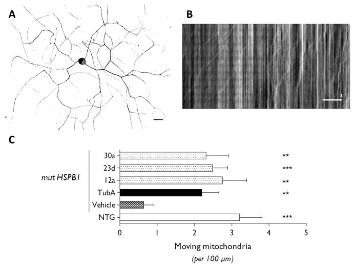
Figure 4.
Axonal transport deficits are rescued by several selective HDAC6 inhibitors. (A) DRG neuron cultured from an adult transgenic HSPB1 mouse is stained for neuronal marker β3-tubulin. Scale bar 50 μm. (B) Axonal transport is measured by tracking the mitochondrial movement within one neurite from each DRG neuron resulting in a kymograph. Vertical lines indicate stationary mitochondria while lines deflecting to the right or left represent anterograde or retrograde moving particles. Horizontal scale bar 20 μm. Vertical scale bar 10 s. (C) Comparing mitochondrial axonal transport in DRG neurons cultured from nontransgenic (NTG) or transgenic (mut HSPB1) mice shows significantly decreased movement in DRG neurons expressing mutant HSPB1. After treatment of the transgenic DRG neurons with 100 nM of the candidate HDAC6 inhibitors, the number of moving mitochondria per 100 μm of neurite was quantified. Similar to the positive control tubastatin A, 12a, 23d, and 30a are able to restore the movement of mitochondria. Graph represents mean values with SEM. Dunn’s multiple comparison test. **p < 0.01, ***p < 0.0001. N = 18–27 for 3–5 mice.

