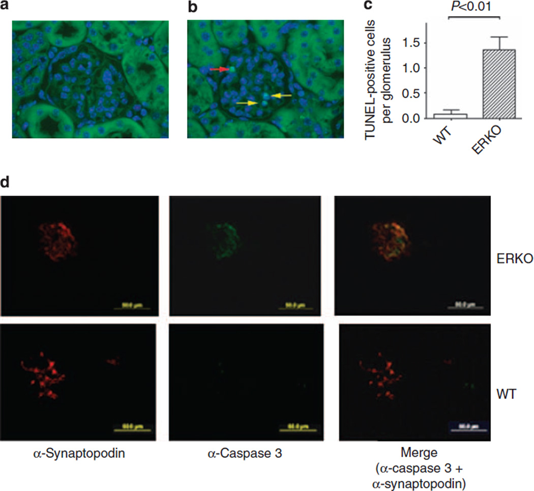
Figure 2. Increased podocyte apoptosis in female αERKO mice.
Podocyte apoptosis in female wild-type (WT) littermates (a) and female αERKO mice (b), as detected by TUNEL assay. Yellow arrows indicate TUNEL-positive cells, whereas the red arrow indicates an artifactual staining of a red blood cell. Results are expressed as percentage of number of TUNEL-positive cells on the total number of glomerular cells and graphed as the mean ± s.e.m. (c). N = 5 mice/group. Original magnifications: × 400. Student’s t-test was performed.
(d) Representative images of caspase 3 immunofluorescence staining and colocalization with synaptopodin expression, used as podocyte marker, in female WT littermates and female αERKO mice. TUNEL, terminal dUTP nick-end labeling.

